


Okay, I admit that my title is meant to be a bit clickbaity. But with clickbaity gusto, I’ll double down and begin by reminding you that Aristotle, who many well-educated people regard as a pretty smart dude, believed that the conscious mind resided in the heart. If you put down all your modern knowledge for a minute, it’s easier to find the idea quite sensible. Consider this utterly imaginary vignette. Aristotle recognizes, through introspection, that when his mind races with emotion, his heart also changes its pace. Thus, Aristotle hypothesizes that the heart is the seat of the soul and decides to do some experiments. He designs an experiment and replicates it in many different types of animals. In this experiment, Aristotle lesions the animal’s heart, and in every case, all behavior indicative of consciousness immediately ends. The result strongly matches some observational data, where accidents caused lesions to the hearts of humans. He also discovers a method to stop the heartbeat through his studies, but only for brief periods. He boldly asks his research assistant to stop his own heart but only for 45 seconds. In this conclusive study, he finds that when his heart is arrested, he immediately becomes unconscious and remains so for the duration of time his heart is stopped. Yet, when his heart begins functioning again, he quickly regains consciousness.
Now we know better than this, and we confidently believe that the nervous system is the seat of the soul. But the fact that such a plausible experiment could lead to such a conclusion should remind us how intimately the vascular system is connected to the brain. I have become obsessed with this connection, and I would love to start my own lab to continue studying it if any universities would hire me (*hint hint*). I want to convince you that cerebrovascular function in the brain is more important than you previously thought. I’ll start by discussing how it’s important for overall brain health. The brain’s vascular system is simultaneously its supply chain distributor and waste management system. Even if you haven’t really thought about it before, I think it is pretty intuitive that if the supply chain breaks down and waste isn’t managed, bad things are going to happen. Then I’ll take things up a notch and talk about how the brain’s vascular function impacts measurements in neuroscience and what that means for how we interpret data. Finally, I’ll turn your brain to mush by taking you deep into the weeds and showing you why I think vascular dynamics might directly organize global brain function and what it would mean for the future of cognitive neuroscience.
Part I
My interest in vascular health started during my graduate school thesis work, which was part of a larger project investigating the role of the vascular system in normal cognitive aging. Our lab was pursuing the hypothesis that dysfunction to the vascular system in the brain contributed to declining mental ability in older adults. This idea is all the rage now, but it was a little less in fashion back then. Art Kramer, a world-renowned cognitive neuroscientist, was doing heroic work at the Beckman Institute, where I was a student, showing that aerobic exercise had a massive effect on predicting cognition in older age. There were a million follow-up questions to be explored. Of these follow-up questions, the most pressing one on everyone’s mind was, “why?” At the time, lots of researchers were sinking their teeth into neurotrophic factors. These molecules support cell growth and survival in the brain and are involved in learning and memory. Their levels increased substantially during aerobic exercise, so many scientists were hypothesizing that they probably explained, at least in part, why aerobic exercise seemed to be so good for cognition. My advisors, Monica Fabiani and Gabriele Gratton, thought that there were probably some significant vascular effects at play, too.
Monica and Gabriele were famous for pioneering the use of the “event-related optical signal” or EROS. The technique measures slight changes in tissue transparency that occur when a region of the brain is active. It offers a favorable trade-off between temporal and spatial resolution for studying the cortex non-invasively. A knock-off version of the technique was even featured on ABC’s one-season-wonder, “Black Box.” However, their technique, “AMORA” (an acronym we could not decode), had far fewer limitations, offering instantaneous, ultra-high-resolution mapping of the whole brain as well as all its activity and pathology.
A well-known obstacle with using EROS is that it is difficult to measure neural activity because the tiny changes in the optical properties of metabolically active tissue were orders of magnitude smaller than changes in the optical properties due to the pulse. Lab members over the years had worked hard to remove this “nuisance” signal from the precious optical measurements that would be used for inferring brain activity.
Monica likes to use the old adage, “one man’s trash is another man’s treasure.” In what I think was a brilliant insight, Monica and Gabriele realized that this “noise” could be repackaged as a “signal” and used for other purposes. This is one of those essential stylistic lessons that I’ll take with me through my scientific career. Scientific discovery is dependent on measurement. Much scientific progress is driven by developing instruments that measure something entirely new or something old with greater precision. But sometimes, it is possible to measure useful things that we haven’t thought to measure yet using devices we already have. An excellent place to look for those potentially “useful” things is by examining the variance in your measurement, even if you were hitherto considering it noise.
Over the following years, our lab explored using different kinds of non-invasive measurements of cerebrovascular health and investigated if those measures predicted brain health and cognition. Using a measure of cerebral blood flow with magnetic resonance imaging (MRI), I conducted an analysis that suggested that age-related declines in blood flow were mediated by fitness level. At the same time, the lab, and especially Chin Hong Tan (now an assistant professor at NTU in Singapore), was developing useful measures from the optical pulse measurement. We found three relatively independent components of the pulse wave: how big it was (amplitude), how fast it was traveling (transit time), and what the shape looked like (the pulse relaxation function or PReFx). These three components predicted different things about an individual’s physiology, anatomy, and cognition.
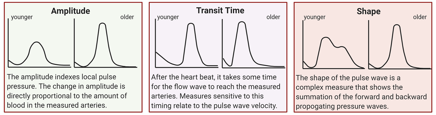
The amplitude of the pulse wave seems to primarily just reflect blood pressure. It doesn’t change much across brain regions, although it is strongly affected by the type of vessels you measure over. If we measure over large arteries, the amplitudes of the pulse wave are higher than if we measure from an area that doesn’t have such large arteries. This is useful because it could be used as a broad proxy for brain anatomy with regions defined by the typical locations of the major surface arteries. Typically, we collect T1-weighted anatomical MRIs to colocalize where our signals are coming from in the brain. Although using the pulse amplitude measures would leave us with a much lower resolution of anatomical features, it is possible to sort of know where you are in the brain in an individual beyond just guessing.
The transit time measures also have some interesting applications. It can be used as a measure of cerebrovascular development in infants, and it can be used to track the flow of the major arteries in the brain.
The most exciting measure is the pulse relaxation function (PReFx) measure that captures the shape of the pulse wave and is sensitive to arterial compliance. Arteries are naturally elastic, which helps absorb some of the pressure from the pulse. Arterial compliance is just the technical term for how much the artery’s volume changes for a given amount of pressure. This PReFx measure predicts cortical volume, white matter lesions, and cognitive performance. The most remarkable thing to me is that it seems to predict regional differences in the brain. For example, PReFx correlates with verbal fluency but not working memory when measured in Broca’s area, an area of the brain implicated in verbal fluency. In contrast, PReFx predicts working memory but not verbal fluency when measured in the dorsolateral prefrontal cortices, an area of the brain implicated in working memory. This kind of double dissociation is often good evidence for the separability of biological systems. That is important because it might predict aspects of health for which no other non-invasive measures exist! There is also some evidence that the PReFx measure is somewhat sensitive to cerebrovascular reactivity, too. Blood flow can be regulated dynamically in the brain to shunt blood around to the areas that need it most. We can see metabolically active regions just by observing changes to the pulse shape. We think that we might be able to use this dynamic attribute to assess the responsiveness of the vessels as well, which could also be an important local predictor of brain health.
In a recent paper led by Tania Kong, we saw tantalizing evidence that there might be a cascade of effects, with changes in vascular health preceding anatomical and functional declines, which finally manifest as cognitive declines. This idea is in line with the cognitive reserve hypothesis that the brain can suffer some variable amount of pathology before any substantial cognitive deficits become measurable. Our analysis just looked at the effect sizes of the correlations, so there are many other possible interpretations of the data. We’re currently following up with longitudinal studies to get a better sense of the causal chain.
In my graduate studies, I also analyzed some more dynamic measures of vascular function. We used breath-holding to make the vessels in the brain vasodilate. When carbon dioxide builds up in the blood, there is a neat protective mechanism in the brain, where some of the small vessels called arterioles globally dilate to increase blood flow. Essentially, the body gets a little suspicious of the environment and sagely says, “let’s prioritize getting oxygen to the brain.” How well the vessels can do this, called vascular reactivity, is an essential feature of vascular function. In my analysis, the amplitude, onset timing, and time-to-recovery of the vasodilatory response varied according to age and fitness. We are currently following up on these observations in ongoing studies.
Part II
These health applications are super exciting, but a bit of a niggling fear started gnawing at me the more I studied the variation in vascular reactivity between individuals and even the variation between regions within individuals. One of our primary measures to infer brain activity in cognitive neuroscience is literally a measure of vascular reactivity. Right now, there is no non-invasive MRI measurement that reliably gives us direct measures of neural activity. Instead, we have a measure sensitive to changes in deoxygenated hemoglobin called the blood-oxygen-level-dependent (BOLD) signal. We’ll take what we can get, but this is not an ideal measure for inferring brain activity. The only reason it works at all is because of a nice physiological quirk, where our brain overcompensates for local increases in metabolic activity by increasing blood flow and overshooting the amount of oxygen it needs. This is great, except that it’s not clear that the overshoot is similar throughout cortical regions or cortical layers, a point that is sometimes acknowledged but usually ignored in practice. Additionally, a lot of the change in deoxygenated hemoglobin occurs in the draining veins. These larger veins drain blood from multiple capillary beds, so it impairs our signal’s spatial resolution by making it harder to identify the precise area the signal comes from. And to top it all off, neurons fire on the scale of milliseconds, but the subsequent hemodynamic response occurs orders of magnitude more slowly, on the order of 4–10s, severely limiting the temporal resolution.
We can learn fascinating things about brain function despite all these limitations by assuming that the relationship between metabolic activity and the hemodynamic response is linear and mostly equivalent between individuals. Thankfully, this linear mapping seems to be more valid than you might expect! The variance between individuals will cancel out in a lot of cognitive experiments, and we learn all sorts of stuff about what the brain does. Unfortunately, sometimes we are primarily interested in group differences, such as different cognitive mechanisms at different ages or in pathology, and that’s when we run into trouble. If we don’t account for systematic differences in vascular physiology, we might create theories about differences in cognitive mechanisms that are actually due to differences in vascular functioning.
An example of this type of group difference research is investigating functional changes in the brain associated with tinnitus or ringing in the ears. Most tinnitus is thought to be “central,” as opposed to “peripheral.” This means that its perception seems to be generated by the brain rather than by the auditory nerve. There are changes to the auditory brain centers thought to be primarily involved in the perception of tinnitus itself. There are also changes in other areas of the brain that correlate to the severity and distress that individuals with tinnitus experience. Some people constantly experience ringing in their ears but get used to it over time in the same way that I don’t really attend to the expressway sounds near my apartment anymore. Unfortunately, other people have a lot of trouble habituating to tinnitus, and it gets much more intrusive and distressing over time. There are two predominant paths of brain research on tinnitus. One of them focuses on understanding what generates the percept in the first place, which is mainly concerned with the primary auditory regions of the brain. The other focuses on understanding why some people do not habituate to it and become so distressed, which appears to involve large areas of interacting brain regions.
In this second category focused on severity, one finding is that subjects with tinnitus have increased brain connectivity between some established networks in the brain compared to research subjects without tinnitus. Activity in a network called the default mode network, which is involved in self-referential processing and is most active at rest, has increased connectivity to networks involved in attention. Another finding is that in subjects with tinnitus, the nodes within the default mode network have decreased connectivity. This finding is typically interpreted within a cognitive framework, using some reverse inference based on past knowledge about what we think these networks do. For example, a researcher may speculate that this activity pattern reflects attention being redirected to the percept of ringing in the ears. This attentional capture intrudes on the usual self-referential thought patterns that occur during rest.
However, it’s essential to know that the correlation between attention and the default mode networks is ordinarily negative. There seems to be some inherent opposition between activity in attention networks and self-referential networks. In the same vein, the correlation of activity of nodes within the default mode network usually is positive. This is important because if our signal-to-noise is different in one group compared to another, we might observe false differences between the groups just because the correlation coefficients in one group are closer to zero than in another group. This is precisely the type of pattern that we observe in tinnitus, so we should be concerned!
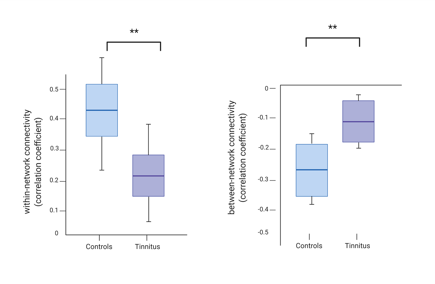
What could cause such a systematic difference in signal-to-noise? Well, at this point, hopefully, my anxiety about differences in vascular physiology has rubbed off on you, and you’re already thinking that differences in vascular reactivity might be responsible for this observation. If the vessels aren’t responding as much to the same amount of metabolic activity, there will be a smaller change in blood oxygenation and thus a smaller BOLD signal.
I didn’t have data on cerebrovascular reactivity measures in tinnitus to look at this directly, but I did have a lovely sample of baseline cerebral blood flow measures in some tinnitus subjects and controls from some of my postdoctoral work with Fatima Husain’s lab. Differences in blood flow do not necessarily reflect differences in cerebrovascular reactivity, but they might suggest it by indicating some vascular health difference. When I looked at this measure, there appeared to be global reductions in cerebral blood flow in tinnitus subjects compared to controls. Even more striking to me, within the tinnitus subjects, blood flow was anti-correlated with tinnitus severity, especially in the places where we tend to see differences in functional connectivity. Here is the link to the peer-reviewed publication.
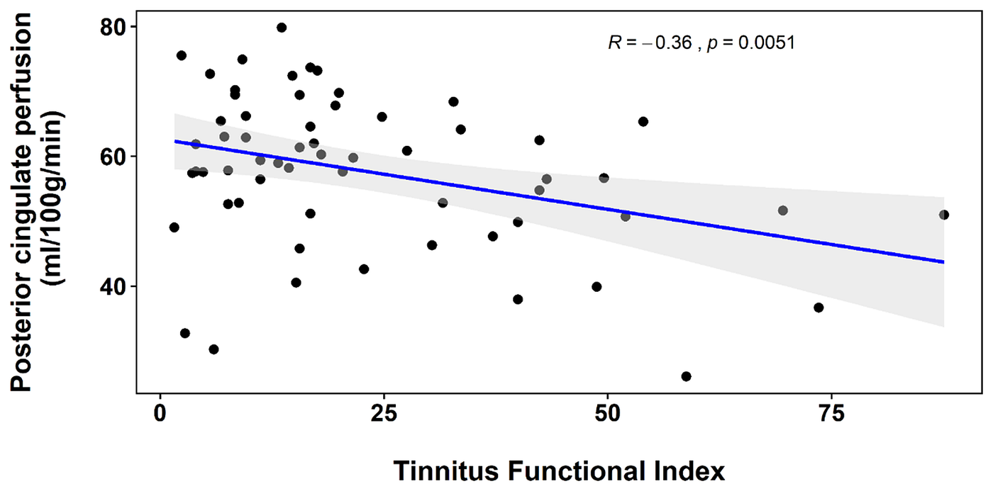
This is not by any means definitive proof that differences in cerebrovascular reactivity cause the functional differences observed in tinnitus. It’s not clear why there would be vascular differences associated with tinnitus, the causal direction between tinnitus and blood flow, or if the blood flow measure really reflects differences in vascular health or instead just reflects different baseline metabolic rates. To me, it’s more of a warning bell that we ought to seriously consider a vascular explanation for these functional differences and do research specifically designed to rule out this possibility.
Neuroscience research on cognitive aging is another research area close to my heart where vascular differences might stymie interpretation. Cerebrovascular function declines with age, so direct comparisons of functional brain mechanisms between age groups are exposed to these same concerns about purely vascular explanations. Two central theoretical ideas in cognitive aging are called “dedifferentiation” and “compensation.” Dedifferentiation proposes that aging is associated with a decrease in the modular structure of functional specialization in the brain, contributing to cognitive decline. Compensation refers to recruiting additional, typically unused neural resources to improve performance, sometimes masking declines in brain health. From a mechanistic point-of-view, if true, both dedifferentiation and neural compensation are extraordinary built-in brain characteristics. They may give us vital clues about the architecture of cognitive processes. Most in silico models of neural networks in artificial intelligence do not exhibit either of these properties. If we ever want to build artificial intelligence that more accurately resembles our brain architecture, we really need to know if these properties are real. Therefore, it is critical to understand whether the data underpinning these theories is affected by cerebrovascular function. Are we measuring actual cognitive mechanisms, or are we just measuring changes in vascular physiology?
Part III
I’ve focused on two main ideas so far. Cerebrovascular mechanisms are crucial because they support cognitive mechanisms by bringing nutrients and oxygen that support neural function. The second idea is that cerebrovascular mechanisms underlie cognitive functional measures, so systematic differences in vascular function can obnubilate interpretations of brain function. A third idea I want to touch on, gaining more traction recently, is that rhythms of vascular dynamics are directly involved in organizing functional activity in the brain.
While working with a team of talented neuroscientists (Fatima Husain and Bahar Shahsavarani) and mathematician/engineers (Yuliy Baryshnikov and Ivan Abraham) to develop a new technique for analyzing fMRI data, called cyclicity analysis, we observed propagating BOLD activity on the order of tens of seconds in the Human Connectome Project data. Our paper is currently under review, but you can read the bioRxiv version here. We found that the BOLD signal activity propagated backward from parietal towards visual areas during rest and the self-reflective part of a social cognition task. In contrast, activity switched directions and propagated forwards from visual areas towards parietal areas during the viewing portion of the social cognition task. I’ve discussed these results in a previous post.
These results align with other labs that have either observed or postulated that unexpected, slow-moving traveling waves of BOLD activity exist in the brain. This slow traveling wave is contentious because it occurs on a much slower time-scale than most oscillatory neural activity or even the hemodynamic response accompanying neural activation. However, if they exist, these “infra-slow” fluctuations in BOLD activity are hypothesized to underlie resting-state functional connectivity.
Resting-state functional connectivity is built upon the observation that activity in certain distributed areas of the brain tends to correlate with other specific areas. The brain appears to self-organize into networks, and those same networks often appear distinct in different types of experimental tasks. The correlations used to determine these networks are traditionally zero-lag correlations, which means that we look at which brain regions are correlated with other brain regions at the same point in time. A key insight is that zero-lag correlations between brain regions would be sensitive to the phase of slow-moving traveling waves.
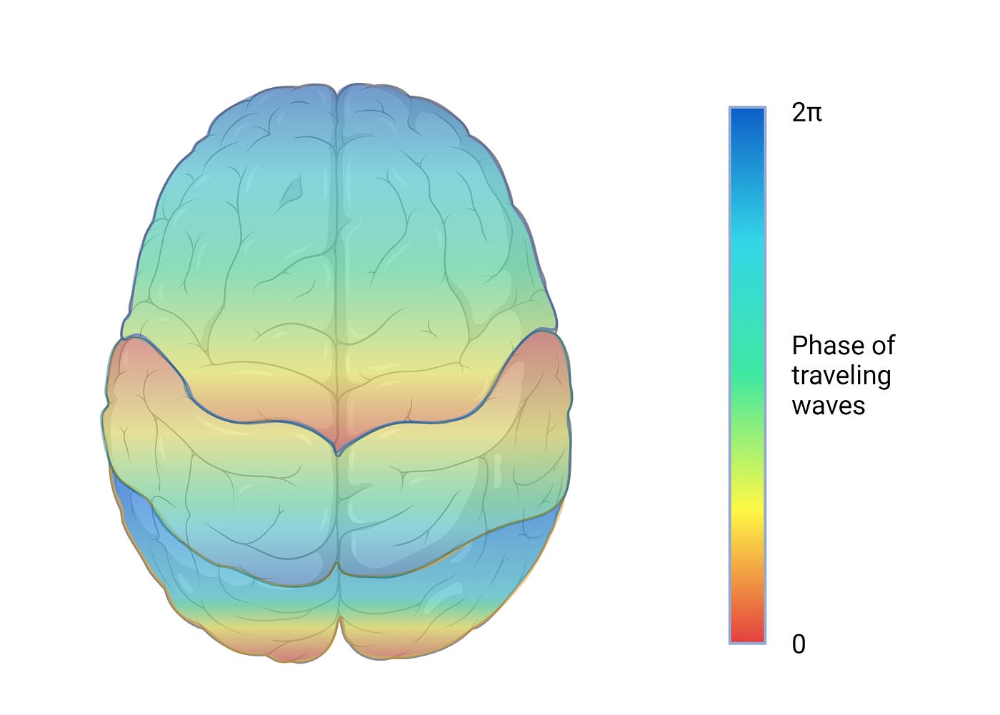
From Marcus Raichle’s lab, Ryan Raut and colleagues recently published data showing that low-frequency BOLD signal fluctuations are coherent with physiological arousal indices, including respiratory variation, heart rate variability, and pupil size. The time it takes for a wave cycle to complete is variable, but the phase of the wave seems locked to the phase of these fluctuations in autonomic arousal. This is pretty amazing! Essentially, they are positing that the functional networks of the brain are generated by slow waves of brain-wide excitability that accompany patterns of autonomic arousal. This is pretty different from the predominant view that functional networks become self-organized according to which areas tend to work together.
What generates these patterns of autonomic arousal? Well, a lot of things could. The autonomic nervous system interacts strongly with many neural systems, breathing, the beating of the heart, the smooth muscle of the vasculature, the stomach and digestion, the bladder, and more. Although it is still unclear what physiology would link autonomic arousal to slow-moving waves in the brain, Raichle’s lab has pointed to this target, autonomic arousal, as the thing to study to better understand brain organization.
However, we still don’t know exactly what is happening when normal functional connectivity is disrupted, such as in aging or certain kinds of pathology. It could be that the autonomic nervous system itself becomes impaired. Alternatively, real functional connections between regions of the brain that normally work together may become impaired. These neural functional connections might normally ride on top of the arousal waves. When they are impaired, it may reduce the signal coming from our measurements, even if the waves of arousal are still intact. Finally, there could be a vascular interaction. The autonomic nervous system also plays a role in controlling the dilation and constriction of the blood vessels. This opening and closing of vessels shifts the supply of blood around the brain. If the effect of the autonomic nervous system on the vessels is blunted or the vasculature cannot properly react to signals to dilate and constrict, then perhaps oxygen and glucose do not get supplied in a timely way. The phase of changes in blood flow might become decoupled from the waves of arousal and impair tissue metabolism.
More and more data are pointing to an unexpected overlap between vascular physiology and functional connectivity. For example, Molly Bright at Northwestern recently found the usual functional connectivity networks within the vascular response to a CO2 inhalation challenge. I told you earlier that a build-up of CO2 should vasodilate the vessels in the brain everywhere in the same way at the same time. “Not so!” says Molly. It looks like the brain’s vascular responsiveness is organized similarly to the functional neural networks. By this, I just mean that the CO2-induced vasodilation within the “default-mode network” is distinguishable from the vasodilation in, say, “attention” networks. We might be tempted to think that the waves of arousal that we see are purely vascular in origin. However, Raichle’s lab and others have observed these global slow waves in electrophysiological data in other animals, suggesting that the vascular response is either parallel to or caused by the changes in neural activity.
My bet is that the interaction of vascular rhythms and functional connectivity will emerge as a significant line of research in the coming years. Here’s my craziest hypothesis: the brain uses these waves of arousal to organize neural activity, and impaired vascular function directly messes up these waves of arousal. To illustrate what I mean by this, imagine that it really is helpful for the dorsal frontal and parietal regions of the brain to coordinate their activity as a network. This coordination is partially facilitated by a wave of arousal that makes neural firing more likely in frontal and parietal regions at the same time. Inflammation in the blood vessels could disrupt autonomic signaling within the brain and de-phase the arousal waves depending on the location of vascular impairment. We already know that inflammation in other areas of the body affects the function of the autonomic nervous system. This is not an easy hypothesis to test because these three levels, autonomic nervous system function, vascular function, and neural function, all interact. I think there are some good starting places to make progress, though. You can experimentally block vasodilation in the small vessels. Does blocking vasodilation affect the slow waves measured with electrophysiology? If it did, it might suggest that cerebral vascular dynamics may themselves be involved in organizing neuronal functional dynamics beyond simply supplying the energetic resource needs of the tissue. If true, it would dramatically alter our view of the role of hemodynamic physiology in brain activity. Along with “supply chain distributor” and “director of waste management,” it would become “project manager.”